

Label-free pathological subtyping of non-small cell lung cancer using deep classification and virtual immunohistochemical staining
npj Digital Medicine, Published online: 03 April 2026; doi:10.1038/s41746-026-02557-x Label-free pathological subtyping of non-small cell lung cancer using deep classification and virtual immunohistochemical staining
References
- Bray, F. et al. Global cancer statistics 2022: GLOBOCAN estimates of incidence and mortality worldwide for 36 cancers in 185 countries. Ca. Cancer J. Clin. 74, 229–263 (2024).
Google Scholar
- Navada, S., Lai, P., Schwartz, A. & Kalemkerian, G. Temporal trends in small cell lung cancer: analysis of the National Surveillance, Epidemiology, and End-Results (SEER) database. J. Clin. Oncol. 24, 7082 (2006).
Google Scholar
- Sher, T., Dy, G. K. & Adjei, A. A. Small cell lung cancer. In Mayo Clinic Proceedings Vol. 83, 355–367 (Elsevier, 2008).
- Kim, H. S., Mitsudomi, T., Soo, R. A. & Cho, B. C. Personalized therapy on the horizon for squamous cell carcinoma of the lung. Lung Cancer 80, 249–255 (2013).
Google Scholar
- Coudray, N. et al. Classification and mutation prediction from non–small cell lung cancer histopathology images using deep learning. Nat. Med. 24, 1559–1567 (2018).
Google Scholar
- Noorbakhsh, J. et al. Deep learning-based cross-classifications reveal conserved spatial behaviors within tumor histological images. Nat. Commun. 11, 6367 (2020).
Google Scholar
- Chen, C.-L. et al. An annotation-free whole-slide training approach to pathological classification of lung cancer types using deep learning. Nat. Commun. 12, 1193 (2021).
Google Scholar
- Yu, K.-H. et al. Classifying non-small cell lung cancer types and transcriptomic subtypes using convolutional neural networks. J. Am. Med. Inform. Assoc. 27, 757–769 (2020).
Google Scholar
- Sadhwani, A. et al. Comparative analysis of machine learning approaches to classify tumor mutation burden in lung adenocarcinoma using histopathology images. Sci. Rep. 11, 16605 (2021).
Google Scholar
- Kanavati, F. et al. A deep learning model for the classification of indeterminate lung carcinoma in biopsy whole slide images. Sci. Rep. 11, 8110 (2021).
Google Scholar
- Chen, J. et al. Automatic lung cancer subtyping using rapid on-site evaluation slides and serum biological markers. Respir. Res. 25, 1–10 (2024).
Google Scholar
- Zhang, M. et al. Decreased green autofluorescence intensity of lung parenchyma is a potential non-invasive diagnostic biomarker for lung cancer. bioRxiv 343533 (2018).
- Wang, N., Liu, Y. & Li, H. An efficient and fast, noninvasive, auto-fluorescence detection method for early-stage oral cancer. IEEE Trans. Instrum. Meas. 71, 1–11 (2022).
Google Scholar
- Waaijer, L. et al. Detection of breast cancer precursor lesions by autofluorescence ductoscopy. Breast Cancer 28, 119–129 (2021).
Google Scholar
- Pu, Y., Wang, W., Yang, Y. & Alfano, R. R. Stokes shift spectroscopic analysis of multifluorophores for human cancer detection in breast and prostate tissues. J. Biomed. Opt. 18, 017005–017005 (2013).
Google Scholar
- Vasanthakumari, P. et al. Discrimination of cancerous from benign pigmented skin lesions based on multispectral autofluorescence lifetime imaging dermoscopy and machine learning. J. Biomed. Opt. 27, 066002–066002 (2022).
Google Scholar
- Becker, W. Fluorescence lifetime imaging–techniques and applications. J. Microsc. 247, 119–136 (2012).
Google Scholar
- Fernandes, S. et al. Fibre-based fluorescence-lifetime imaging microscopy: a real-time biopsy guidance tool for suspected lung cancer. Transl. Lung Cancer Res. 13, 355 (2024).
Google Scholar
- Adams, A. C. et al. Fibre-optic based exploration of lung cancer autofluorescence using spectral fluorescence lifetime. Biomed. Opt. Express 15, 1132–1147 (2024).
Google Scholar
- Zang, Z. et al. Fast analysis of time-domain fluorescence lifetime imaging via extreme learning machine. Sensors 22, 3758 (2022).
Google Scholar
- Sorrells, J. E. et al. Real-time pixelwise phasor analysis for video-rate two-photon fluorescence lifetime imaging microscopy. Biomed. Opt. Express 12, 4003–4019 (2021).
Google Scholar
- Luo, T., Lu, Y., Liu, S., Lin, D. & Qu, J. Phasor–FLIM as a Screening tool for the differential diagnosis of actinic keratosis, Bowen’s disease, and basal cell carcinoma. Anal. Chem. 89, 8104–8111 (2017).
Google Scholar
- Walsh, A. J. et al. Classification of T-cell activation via autofluorescence lifetime imaging. Nat. Biomed. Eng. 5, 77–88 (2021).
Google Scholar
- Hu, L., Ter Hofstede, B., Sharma, D., Zhao, F. & Walsh, A. J. Comparison of phasor analysis and biexponential decay curve fitting of autofluorescence lifetime imaging data for machine learning prediction of cellular phenotypes. Front. Bioinforma. 3, 1210157 (2023).
Google Scholar
- Alfonso-García, A. et al. Label-free identification of macrophage phenotype by fluorescence lifetime imaging microscopy. J. Biomed. Opt. 21, 046005 (2016).
Google Scholar
- Wang, Q. et al. Deep learning in ex-vivo lung cancer discrimination using fluorescence lifetime endomicroscopic images. In 2020 42nd annual international conference of the IEEE Engineering in Medicine & Biology Society (EMBC) 1891–1894 (IEEE, 2020).
- Wang, Q., Vallejo, M. & Hopgood, J. Fluorescence lifetime endomicroscopic image-based ex-vivo human lung cancer differentiation using machine learning. Authorea Prepr. (2023).
- Wang, Q. et al. A layer-level multi-scale architecture for lung cancer classification with fluorescence lifetime imaging endomicroscopy. Neural Comput. Appl. 34, 18881–18894 (2022).
Google Scholar
- Wang, Q. et al. Deep learning-based virtual H& E staining from label-free autofluorescence lifetime images. Npj Imaging 2, 17 (2024).
Google Scholar
- Bai, B. et al. Deep learning-enabled virtual histological staining of biological samples. Light Sci. Appl. 12, 57 (2023).
Google Scholar
- Kreiss, L. et al. Digital staining in optical microscopy using deep learning-a review. PhotoniX 4, 34 (2023).
Google Scholar
- Rivenson, Y. et al. Virtual histological staining of unlabelled tissue-autofluorescence images via deep learning. Nat. Biomed. Eng. 3, 466–477 (2019).
Google Scholar
- Zhang, Y. et al. Digital synthesis of histological stains using micro-structured and multiplexed virtual staining of label-free tissue. Light Sci. Appl. 9, 78 (2020).
Google Scholar
- Li, X. et al. Unsupervised content-preserving transformation for optical microscopy. Light Sci. Appl. 10, 44 (2021).
Google Scholar
- DoanNgan, B., Angus, D., Sung, L., & others. Label-free virtual HER2 immunohistochemical staining of breast tissue using deep learning. BME Front. (2022).
- Zhang, G. et al. Image-to-images translation for multiple virtual histological staining of unlabeled human carotid atherosclerotic tissue. Mol. Imaging Biol. 1–11 (2022).
- Borhani, N., Bower, A. J., Boppart, S. A. & Psaltis, D. Digital staining through the application of deep neural networks to multi-modal multi-photon microscopy. Biomed. Opt. Express 10, 1339–1350 (2019).
Google Scholar
- Kang, L., Li, X., Zhang, Y. & Wong, T. T. Deep learning enables ultraviolet photoacoustic microscopy based histological imaging with near real-time virtual staining. Photoacoustics 25, 100308 (2022).
Google Scholar
- Cao, R. et al. Label-free intraoperative histology of bone tissue via deep-learning-assisted ultraviolet photoacoustic microscopy. Nat. Biomed. Eng. 7, 124–134 (2023).
Google Scholar
- Levy, J. J., Jackson, C. R., Sriharan, A., Christensen, B. C. & Vaickus, L. J. Preliminary evaluation of the utility of deep generative histopathology image translation at a mid-sized NCI cancer center. BioRxiv 2020–01 (2020).
- Hong, Y. et al. Deep learning-based virtual cytokeratin staining of gastric carcinomas to measure tumor–stroma ratio. Sci. Rep. 11, 19255 (2021).
Google Scholar
- Lahiani, A., Klaman, I., Navab, N., Albarqouni, S. & Klaiman, E. Seamless virtual whole slide image synthesis and validation using perceptual embedding consistency. IEEE J. Biomed. Health Inform. 25, 403–411 (2020).
Google Scholar
- Zhang, R. et al. MVFStain: multiple virtual functional stain histopathology images generation based on specific domain mapping. Med. Image Anal. 80, 102520 (2022).
Google Scholar
- Lin, Y. et al. Unpaired multi-domain stain transfer for kidney histopathological images. Proc. AAAI Conf. Artif. Intell. 36, 1630–1637 (2022).
Google Scholar
- Pati, P. et al. Accelerating histopathology workflows with generative AI-based virtually multiplexed tumour profiling. Nat. Mach. Intell. 6, 1077–1093 (2024).
Google Scholar
- Moldvay, J. et al. The role of TTF-1 in differentiating primary and metastatic lung adenocarcinomas. Pathol. Oncol. Res. 10, 85–88 (2004).
Google Scholar
- Affandi, K. A., Tizen, N. M. S., Mustangin, M. & Zin, R. R. M. R. M. p40 immunohistochemistry is an excellent marker in primary lung squamous cell carcinoma. J. Pathol. Transl. Med. 52, 283–289 (2018).
Google Scholar
- Yatabe, Y. et al. Best practices recommendations for diagnostic immunohistochemistry in lung cancer. J. Thorac. Oncol. 14, 377–407 (2019).
Google Scholar
- Goodwin, J. et al. The distinct metabolic phenotype of lung squamous cell carcinoma defines selective vulnerability to glycolytic inhibition. Nat. Commun. 8, 15503 (2017).
Google Scholar
- Song, Q. et al. Proteomic analysis reveals key differences between squamous cell carcinomas and adenocarcinomas across multiple tissues. Nat. Commun. 13, 4167 (2022).
Google Scholar
- Chattopadhay, A., Sarkar, A., Howlader, P. & Balasubramanian, V. N. Grad-cam + +: Generalized gradient-based visual explanations for deep convolutional networks. in 2018 IEEE winter conference on applications of computer vision (WACV) 839–847 (IEEE, 2018).
- Cai, T. T. & Ma, R. Theoretical foundations of t-SNE for visualizing high-dimensional clustered data. J. Mach. Learn. Res. 23, 1–54 (2022).
Google Scholar
- He, K., Zhang, X., Ren, S. & Sun, J. Deep Residual Learning for Image Recognition. In 2016 IEEE Conference on Computer Vision and Pattern Recognition (CVPR) 770–778 (2016). https://doi.org/10.1109/CVPR.2016.90.
- Tan, M. EfficientNet: Rethinking model scaling for convolutional neural networks. ArXiv Prepr. ArXiv190511946 (2019).
- Huang, G., Liu, Z., Van Der Maaten, L. & Weinberger, K. Q. Densely connected convolutional networks. In Proceedings of the IEEE conference on computer vision and pattern recognition 4700–4708 (2017).
- WHO Classification of Tumours Editorial Board. Thoracic Tumours. WHO Classification of Tumours, 5th Edition, Volume 5. (International Agency for Research on Cancer, 2021).
- Zhang, M. et al. Decreased green autofluorescence of lung parenchyma is a biomarker for lung cancer tissues. J. Biophotonics 15, e202200072 (2022).
Google Scholar
- Vorontsov, E. et al. A foundation model for clinical-grade computational pathology and rare cancers detection. Nat. Med. 30, 1–12 (2024).
- Wang, X. et al. A pathology foundation model for cancer diagnosis and prognosis prediction. Nature 634, 970–978 (2024).
Google Scholar
- Vaswani, A. Attention is all you need. Adv. Neural Inf. Process. Syst. (2017).
- Liu, Z. et al. A convnet for the 2020s. In Proceedings of the IEEE/CVF conference on computer vision and pattern recognition 11976–11986 (IEEE, 2022).
- Bai, B. et al. Label-free virtual HER2 immunohistochemical staining of breast tissue using deep learning. BME Front 2022, 9786242 (2022).
Google Scholar
- Dalmaz, O. & Yurt, M. & Çukur, T. ResViT: Residual vision transformers for multimodal medical image synthesis. IEEE Trans. Med. Imaging 41, 2598–2614 (2022).
Google Scholar
- Zhang, Y. et al. Super-resolved virtual staining of label-free tissue using diffusion models. ArXiv Prepr. ArXiv241020073 (2024).
- Kataria, T., Knudsen, B. & Elhabian, S. Y. StainDiffuser: multitask dual diffusion model for virtual staining. ArXiv Prepr. ArXiv240311340 (2024).
- Preibisch, S., Saalfeld, S. & Tomancak, P. Globally optimal stitching of tiled 3D microscopic image acquisitions. Bioinformatics 25, 1463–1465 (2009).
Google Scholar
- Isola, P., Zhu, J.-Y., Zhou, T. & Efros, A. A. Image-to-image translation with conditional adversarial networks. in Proceedings of the IEEE conference on computer vision and pattern recognition 1125–1134 (IEEE, 2017).
- Wightman, R. PyTorch Image Models. GitHub repository https://doi.org/10.5281/zenodo.4414861 (2019).
- Wang, Z., Bovik, A. C., Sheikh, H. R. & Simoncelli, E. P. Image quality assessment: from error visibility to structural similarity. IEEE Trans. Image Process. 13, 600–612 (2004).
Google Scholar
- Gatys, L. A. A neural algorithm of artistic style. ArXiv Prepr. ArXiv150806576 (2015).
Download references
Sign in to highlight and annotate this article
Conversation starters
Daily AI Digest
Get the top 5 AI stories delivered to your inbox every morning.
More about
published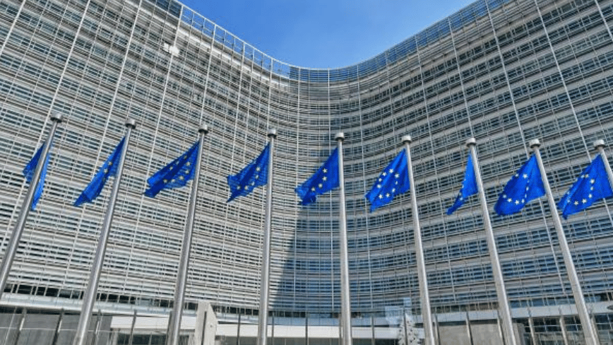
Hackers breached the European Commission by poisoning the security tool it used to protect itself
CERT-EU has attributed a major data breach at the European Commission to cybercrime group TeamPCP, which exploited a supply chain attack on the open-source security tool Trivy to steal 92 GB of compressed data from the Commission’s AWS infrastructure. The notorious ShinyHunters gang then published the data, which included emails and personal details from up [ ] This story continues at The Next Web

HoloTrauma 3X Triadic AI Co reasoning for robot assisted emergency maxillofacial reconstruction
npj Digital Medicine, Published online: 04 April 2026; doi:10.1038/s41746-026-02573-x HoloTrauma 3X Triadic AI Co reasoning for robot assisted emergency maxillofacial reconstruction
Knowledge Map
Connected Articles — Knowledge Graph
This article is connected to other articles through shared AI topics and tags.
More in Research Papers
New Rowhammer attack can grant kernel-level control on Nvidia workstation GPUs
A study from researchers at UNC Chapel Hill and Georgia Tech shows that GDDR6-based Rowhammer attacks can grant kernel-level access to Linux systems equipped with GPUs based on Nvidia's Ampere and Ada Lovelace architectures. The vulnerability appears significantly more severe than what was outlined in a paper last year. Read Entire Article
![[D] ICML Reviewer Acknowledgement](https://d2xsxph8kpxj0f.cloudfront.net/310419663032563854/konzwo8nGf8Z4uZsMefwMr/default-img-matrix-rain-CvjLrWJiXfamUnvj5xT9J9.webp)
[D] ICML Reviewer Acknowledgement
Hi, I'm a little confused about ICML discussion period Does the period for reviewer acknowledging responses have already ended? One of the four reviewers did not present any answer to a paper of mine. Do you know if the reviewer can still change their score before April 7th? There is a reviewer comment that I will answer on Monday. Will the reviewer be able to update the score after seeing my answer? Thanks! submitted by /u/Massive_Horror9038 [link] [comments]

Considerations for growing the pie
Recently some friends and I were comparing growing the pie interventions to an increasing our friends' share of the pie intervention, and at first we mostly missed some general considerations against the latter type. 1. Decision-theoretic considerations The world is full of people with different values working towards their own ends; each of them can choose to use their resources to increase the total size of the pie or to increase their share of the pie. All of them would significantly prefer a world in which resources were used to increase the size of the pie, and this leads to a number [of] compelling justifications for each individual to cooperate. . . . by increasing the size of the pie we create a world which is better for people on average, and from behind the veil of ignorance we s

Discussion
Sign in to join the discussion
No comments yet — be the first to share your thoughts!